Researchers are working to replace gold standard of breast tissue scanning with a Photoacoustic Methodology
Orange County, CA - June 7th 2017 - Annually, around 180,000 women in the United States undergo surgery to remove cancerous breast tissue while simultaneously trying to preserve as much healthy tissue as possible. There is no method currently available that can show surgeons how much cancerous tissue is left while operating. The method considered to be the gold standard of scanning takes multiple days to analyze post-op images of exacted tissue. As a result, a quarter of the women who underwent surgery later receive word of needing to endure a second procedure due to leftover cancerous tissue.
Researchers at the Washington University School of Medicine in St. Louis and the California Institute of Technology in Pasadena have reported they are developing a new method. By implementing photoacoustic imaging to scan a retrieved tumor sample, results are delivered in a timely fashion unlike current methodologies. However, researchers concede that more work is to be done before this method is quick enough to use in an operation.
Published in the journal Science Advances, Deborah Novak, MD, PhD- associate professor of medicine, pathology, and immunology- and a co-senior author on this study, states, “This is a proof of concept that we can use photoacoustic imaging on breast tissue and get images back that look similar to traditional staining methods without any sort of tissue processing.” Given a little more time, researchers believe the scanning of specimens can be brought down to 10 minutes. Given this speed the procedure will be more than fast enough to be done during a surgery while patients are sedated. A medical advancement of this area and caliber has yet to be seen since the mid-20th century.
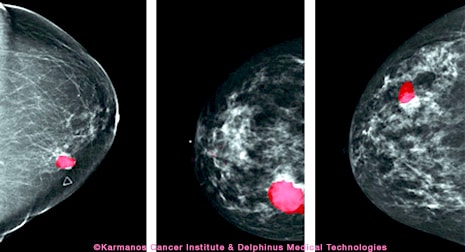 Currently, to examine if any remaining cancerous tissue needs to be extracted, a pathologist slices a tissue sample and stains it with dye to inspect for margins of malignant cells under a microscope. What pathologists expect to see is an ample amount of normal cells surrounding the treated area, suggesting all the malignant cells were successfully removed. If there are remaining malignant cells close to the perimeter of the healthy tissue, the pathologist will presume cancerous tissue was left behind in the patient. This process is known as inspecting a “frozen section.” To further complicate things, inspecting a frozen section is more difficult on breast tissue because the process isn’t as efficient when examining fatty tissue specimens. To test the abilities of photoacoustic imaging under such constructs, researchers scanned 3 cancer patients’ tumor slices in comparison to three other cancerous specimens stained in the traditional fashion. The photoacoustic image matched the stained samples in all aspects of testing. Having established the accuracy in photoacoustic technique, researchers are prioritizing the reduction of overall time needed for testing.
“One day we think we’ll be able to take a specimen straight from the patient, plop it into the machine in the operating room and know in minutes whether we’ve gotten [the entire] tumor out or not,” said Rebecca Aft, MD, PhD, professor of surgery, and the other co-author of the study. “That’s the goal.”
Contact Ampronix:

Email: info@ampronix.com
International Sales: +1 949-273-8000
Domestic Sales: 1800-400-7972 for US and Canada
Follow Us:
Share This Article:


View our Product Catalog Online Here
About Ampronix
Ampronix is a renowned authorized master distributor of the medical industry's top brands as well as a world class manufacturer of innovative technology. Since 1982, Ampronix has been dedicated to meeting the growing needs of the medical community with its extensive product knowledge, outstanding service, and state-of-the-art repair facility. Ampronix prides itself on its ability to offer tailored, one-stop solutions at a faster and more cost effective rate than other manufacturers.
Ampronix is an ISO & ANSI/ESD certified facility. To learn more go here.
![]()
Researchers are working to replace gold standard of breast tissue scanning with a Photoacoustic Methodology Orange County, CA – June 7th 2017 – Annually, around 180,000 women in the United States undergo surgery to remove cancerous breast tissue while simultaneously trying to preserve as much healthy tissue as possible. There is no method currently available that […]