Comparing new and old methods of diagnosis to confirm the accuracy of results more quickly
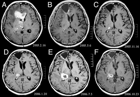
Orange County, CA - February 9th 2017 - Neurosurgeons and pathologists at Michigan Medicine have recently trialed a new method of surgical pathology to improve operations of patients suffering with brain tumors. The technique, called stimulated Raman histology (SRH), is the first of its kind and is able to improve speed, safety, and diagnostic efficiency of the surgical procedure. Researchers believe if the technique is applied universally, it could revise the momentum and framework of the operation.
The study tested tissue samples from 101 neurosurgical patients using both conventional methods and the new SRH method. Assessment found that both techniques produced similarly accurate results, but that the new method was done more rapidly.
At present, when trying to confirm a diagnosis during an operation, surgeons must send tissue samples to the pathology lab to process, section, stain, mount, and interpret the results. This procedure can take up to 30 to 40 minutes where, in the middle of an operation, the entire surgical team is at a stand still awaiting results. The new technique can turn 30 minutes into three.
"It's very similar to what we currently do in our intraoperative diagnosis, with the exception that the tissue is fresh, [and] has not been processed or stained," said Sandra Camelo-Piragua, M.D., senior author and assistant professor of pathology at the U-M Medical School.
The technology behind SRH is called stimulated Raman scattering microscopy and was developed in 2008. At the time of its creation the lasers used in the procedure were considered harmful and not appropriate for operating room use, but after more than a year of testing at the University of Michigan, a clinical version was created for research. The fiber-laser-based microscope is mounted on a medical cart that is able to plug directly into a wall socket.
The SRH uses virtual coloring to distinguish between the cellular and architectural characteristics of brain tumors and normal tissue, similar to how traditional staining is carried out. Daniel A. Orringer, M.D., first author and assistant professor of neurosurgery at the University of Michigan Medical School, and his team are also using artificial intelligence to teach a computer to diagnose based on SRH images. Together they were able to build a machine learning process with a 90 percent accuracy rate in detecting tumor subtypes.
The new clinical version of the stimulated Raman scattering microscopy has yet to undergo clinical trials, but Dr. Orringer believes the technology could be of use to facilities where an expert neuropathologists isn’t available.
Contact Ampronix:
Email: info@ampronix.com
International Sales: +1 949-273-8000
Domestic Sales: 1800-400-7972 for US and Canada
Follow Us:
Share This Article:


View our Product Catalog Online Here
About Ampronix
Ampronix is a renowned authorized master distributor of the medical industry's top brands as well as a world-class manufacturer of innovative technology. Since 1982, Ampronix has been dedicated to meeting the growing needs of the medical community with its extensive product knowledge, outstanding service, and state-of-the-art repair facility. Ampronix prides itself on its ability to offer tailored, one-stop solutions at a faster and more cost-effective rate than other manufacturers. Ampronix is an ISO & ANSI/ESD certified facility. To learn more go here.
![]()
Comparing new and old methods of diagnosis to confirm the accuracy of results more quickly Orange County, CA – February 9th 2017 – Neurosurgeons and pathologists at Michigan Medicine have recently trialed a new method of surgical pathology to improve operations of patients suffering with brain tumors. The technique, called stimulated Raman histology (SRH), is the first […]